Hella A. Bolck, Ede Migh, Andras Kriston, Natalia Zajac, Susanne Kreutzer, Tiberiu Totu, Peter Leary, Ferenc Kovacs, Dorothea Rutishauser, Sibylle Pfammatter, Jonas Grossmann, Cassandra Litchfield, Marija Buljan, Niels J. Rupp, Peter Horvath, Holger Moch
Bolck et al. present a novel “Deep Visual Multi-Omics” workflow to characterize the molecular features behind different morphological grades of clear cell renal cell carcinoma (ccRCC). Our Biological Image Analysis Software (BIAS) was a central component of this approach, using artificial intelligence to classify individual cancer cells from pathology slides, which enabled their targeted isolation for deep molecular analysis. This work, available as a preprint, identifies unique molecular signatures of highly aggressive rhabdoid cancer cells, providing a basis for developing new combination therapies.
Linking Cellular Morphology to Molecular Function
Clear cell renal cell carcinoma (ccRCC) is characterized by significant heterogeneity; a single tumor can contain a mixture of cells with different appearances and behaviors. This diversity, particularly the presence of high-grade “rhabdoid” cells, is associated with poor patient outcomes. For a long time, the relationship between a cell’s morphology and its underlying molecular state has been poorly understood. Clarifying this link is essential for developing more effective, targeted treatments.
To address this, researchers developed an integrated workflow that establishes a direct link between digital pathology and molecular biology. This method involves a multi-step process:
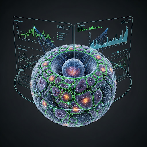
- AI-Powered Digital Pathology with BIAS: The process begins with standard, H&E-stained pathology slides. These are digitized into high-resolution images and analyzed using our BIAS software. Employing deep learning models, BIAS automatically segments and classifies tens of thousands of individual cells, distinguishing between normal kidney tissue and different tumor grades (low-grade, high-grade, and rhabdoid) with high accuracy. This automated and precise classification is a critical first step that informs the subsequent molecular analysis.
- Precision Single-Cell Isolation: Based on the classification map created by BIAS, a laser microdissection system is used to isolate specific cells of interest. For this study, the team collected two sets of 1,000 cells from each morphological class (low-grade, high-grade, rhabdoid, and normal) from five different patients.
- Deep Multi-Omics Profiling: These pure populations of morphologically defined cells were then subjected to sensitive transcriptomic (mRNA) and proteomic (protein) analysis. This provided a comprehensive view of the active genes and proteins in each cell type.
The analysis revealed a clear pattern of increasing molecular dysregulation that correlated directly with the increasing histopathological grade. The rhabdoid cells, the most aggressive type, exhibited a distinct molecular profile. Their machinery for cell proliferation, driven by the transcription factor FOXM1, was highly active. Concurrently, their cell-adhesion programs were disrupted, which is consistent with their discohesive growth pattern and likely contributes to their metastatic potential.
Importantly, the study identified a specific immune evasion mechanism employed by rhabdoid cells. Although these tumor areas were often infiltrated with tumor-fighting CD8+ T-cells, the rhabdoid cells were actively creating an immunosuppressive environment. They upregulated the known immune checkpoint PD-L1 and also novel immunomodulatory factors such as CD38 and ITGB2, likely contributing to the functional exhaustion of the T-cells.
This research provides a rationale for developing new therapeutic strategies. By identifying the specific molecular mechanisms of the most aggressive cancer cells, we can devise more rational combination therapies. For patients with rhabdoid ccRCC, this could mean pairing immune checkpoint inhibitors with agents that target FOXM1, CD38, or other key nodes in the rhabdoid-specific networks, with the goal of restoring an effective anti-tumor immune response.