Akos Diosdi, Timea Toth, Maria Harmati, Istvan Grexa, Bálint Schrettner, Nora Hapek, Ferenc Kovacs, Andras Kriston, Krisztina Buzas, Francesco Pampaloni, Filippo Piccinini, Peter Horvath
Diosdi et al. introduce HCS-3DX, a next-generation, AI-driven system designed to overcome the challenges of high-content screening in complex 3D cell cultures. This integrated platform, developed in-house at Single-Cell Technologies, leverages our Biological Image Analysis Software (BIAS) to enable precise, single-cell level analysis of spheroids and organoids, paving the way for more accurate drug discovery and personalized medicine.
Advancing High-Content Screening into the Third Dimension
For years, drug discovery and cell biology have relied on studying cells grown in flat, 2D layers on plastic dishes. While convenient, this method fails to capture the complex interactions and architecture of cells within a living organism. Three-dimensional cell cultures, such as spheroids and organoids, offer a much more physiologically relevant model, but their complexity has posed significant challenges for automated, high-throughput analysis.
The HCS-3DX system, developed by our team at Single-Cell Technologies, addresses these challenges head-on. It is a complete, end-to-end solution that integrates several innovations:
- AI-Driven Object Selection: A key bottleneck in 3D screening is the manual and subjective process of selecting which spheroids or organoids are suitable for imaging. HCS-3DX automates this with an AI-based prescreening step. The system rapidly scans all wells, identifies all 3D objects, and uses machine learning to select only the most viable and representative candidates for high-resolution imaging, saving significant time and resources.
- Optimized Imaging Hardware: To achieve the best possible images deep within 3D structures, the system utilizes proprietary optical-bottom plates. These plates are designed to minimize optical distortions, allowing for clearer, higher-resolution images to be captured by the spinning-disk confocal microscope, which is essential for detailed single-cell analysis.
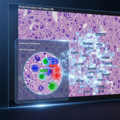
- Advanced Image Analysis with BIAS: At the heart of the HCS-3DX system is our BIAS software. The high-resolution 3D image stacks are processed using sophisticated deep-learning algorithms within BIAS. This allows for the accurate segmentation of individual nuclei even in densely packed spheroids. From these segmentations, the software can extract a rich set of features for each cell, such as its location within the spheroid, its morphology, and the intensity of fluorescent markers.
This ability to perform detailed analysis at the single-cell level within a 3D context is a significant advance for the field. It allows researchers to understand how a potential drug affects not just the whole spheroid, but also different cell populations within it. The authors demonstrate the power of the HCS-3DX system by showing how it can quantify the dose-dependent effects of an anti-cancer drug on different cell cycle phases, revealing spatial patterns of drug response within the spheroid.
By creating a robust, automated, and highly quantitative platform for 3D cell culture analysis, the HCS-3DX system opens up new possibilities for more predictive preclinical drug screening and the development of personalized cancer therapies based on patient-derived organoids. We believe this technology represents a critical step forward in bridging the gap between laboratory models and clinical reality.