Conclusion of the first SCT Single-cell Isolation Workshop
What an amazing two days we had at our first ever multi-day Single-cell Isolation Workshop last week! Our Single Cell Workshop has just wrapped up, and we couldn’t be prouder.We […]
What an amazing two days we had at our first ever multi-day Single-cell Isolation Workshop last week! Our Single Cell Workshop has just wrapped up, and we couldn’t be prouder.We […]
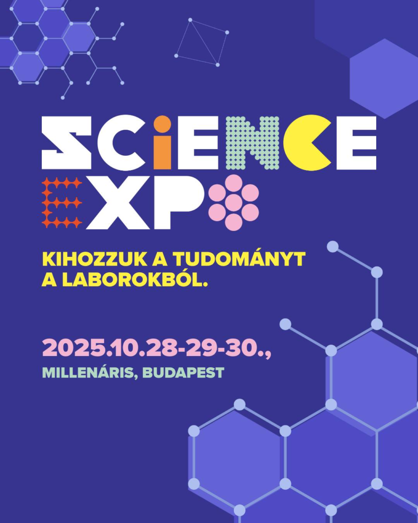
We are excited to announce that Single-Cell Technologies (SCT) will be present at the 2025 Science Expo organized by the Hungarian Innovation Agency and HUN-REN, taking place at Millenáris, Budapest,

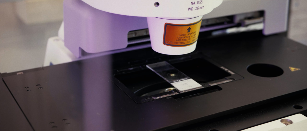
Szferle et al. have developed an AI-based pathology workflow to objectively quantify and predict heart transplant rejection from biopsy images . This study, published in the Hungarian medical journal Orvosi Hetilap, centered on optimizing and applying our Biological Image Analysis Software (BIAS) to automatically identify cells and measure key morphological parameters that indicate rejection . The research, involving team members from our company, Single-Cell Technologies, successfully demonstrated that BIAS can quantify parameters like lymphocyte density and proximity to heart muscle cells, which strongly correlate with the severity of graft rejection, offering a new and powerful quantitative tool for pathologists .
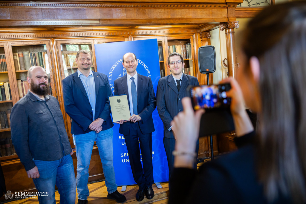
We are proud to share that this summer, our collaborative research was honored with the Markusovszky Lajos Prize, awarded by Orvosi Hetilap (Hungarian Medical Journal). The prize recognized the article:
We are excited to announce SCT’s first ever multi-day Single-cell Isolation Workshop to be held at the Single Cell Centre, Szeged, Hungary on the November 10-11, 2025.
Registration is free but places are limited, and will remain open until the end of September 2025.
For details and registration, please visit the event webpage.
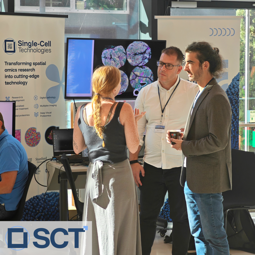
Last week, we had the privilege to join the 6th European Symposium on Single Cell Proteomics (ESCP) in Vienna. It was an exciting double milestone for us as we had
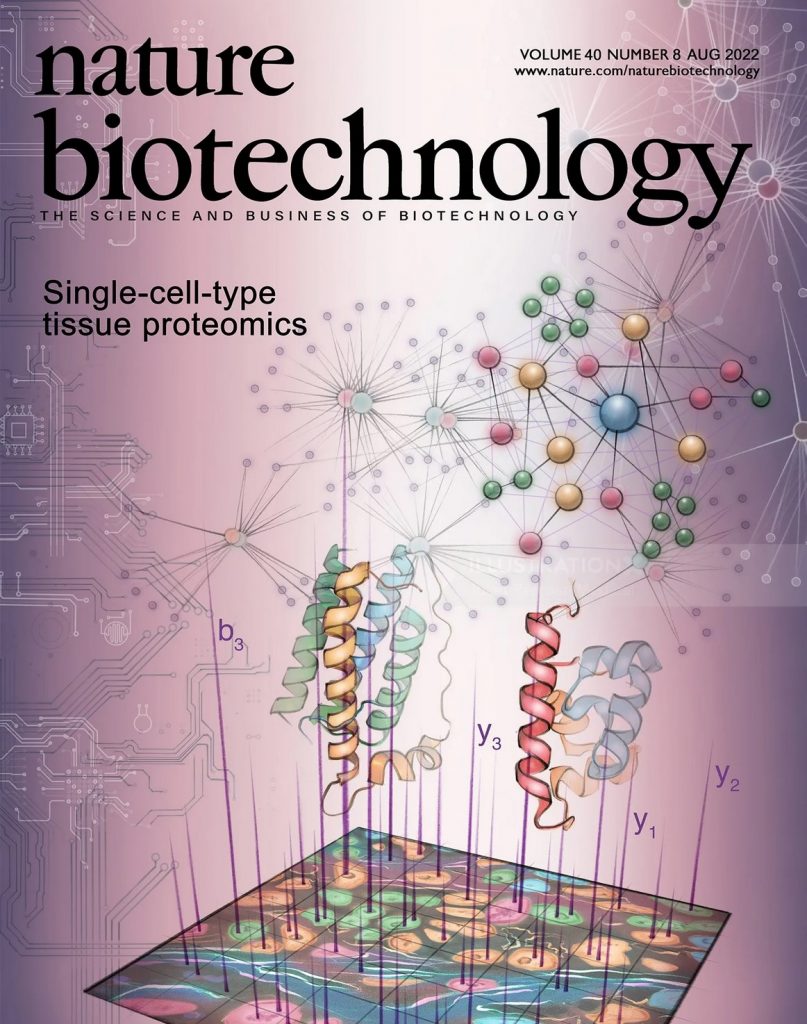
Mund et al. have developed a revolutionary method called Deep Visual Proteomics (DVP), which for the first time, allows for the analysis of thousands of proteins from single cells while keeping their original location in the tissue intact. Our Biological Image Analysis Software (BIAS) was a key component of this research, providing the powerful AI-driven image analysis needed to identify and classify cells for proteomic analysis. This groundbreaking work, published in Nature Biotechnology, opens up new avenues for understanding the molecular details of diseases like cancer.